

Serum albumins are important in regulating blood volume by maintaining the oncotic pressure (also known as colloid osmotic pressure) of the blood compartment, bovine serum albumin (cattle serum albumin) or BSA, often used in medical and molecular biology laboratories. Serum albumin is the main protein of human blood plasma. It binds water, cations (such as Ca2+, Na+ and K+), fatty acids, hormones, bilirubin, Thyroxin (T4) and pharmaceuticals (including barbiturates), its main function is to regulate the oncotic pressure of blood. Alpha and beta globulins function as enzymes and proteins that transport compounds in the body. Gamma globulins act as the antibody defence against antigen invasion (Aarons and Rowland, 1981; Aggeler et al., 1967) Gamma globulins are manufactured in cells of the immune system known as lymphocytes and plasma cell, Blood plasma, which is usually about 4% of the body weight, consists of many constituents including H2O, nutrients and metabolites, proteins, various hormones and electrolytes (Aarons and Rowland, 1981). The normal total plasma protein concentration ranges from 5.5 to 8.9 g/dL in domestic-animal species, and it is a complex mixture including simple proteins, glycoproteins and various lipoprotein housands of antibodies (immunoglobulins) are also present in plasma, although under normal conditions the amount of any one is usually quite low.
Abstract
Share on Mobile
Scan to access on mobile
Keywords
Monoclonal Gammopathy
Plasma Proteins Electrophoresis
Polyclonal Gammopathy
Serum Albumins Serum Proteins
INTRODUCTION
Plasma proteins are important complementary constituents in the diagnosis of gastrointestinal, hepatic, renal, or infectious diseases. Determination of plasma proteins image1seldom leads to a specific diagnosis (e.g., monoclonal gammopathies) but will help the clinician to evaluate the nature, severity, and progress of a disease (Table 1). Blood plasma contains 8% solids, which has 7% albumin. The different plasma proteins are albumins, globulins, and fibrinogen. Usually, total plasma proteins are 6 to 8 g / 100 mL of blood. The proteins present in human blood plasma are a mixture of simple proteins, glycoproteins, lipoproteins (Banfield et al., 1983). And other conjugated proteins called “Plasma Protein”. The Functions of Plasma Proteins; Plasma proteins are a crucial component of blood that perform many functions in the human body. They are produced in the liver and circulate in the blood, which is vital in maintaining our overall health. Plasma protein is vital in our body. Here are the primary functions.
Table 1: Plasma proteins and clinical significance case study 1.
| Protein molecular weight (g/mol) | Concentration in serum (g/L) | Half-life (days) | Function | Common causes for increased (↑) or decreased (↓) concentrations |
|---|---|---|---|---|
| Transthyretin (Prealbumin) 54,000 | 0.2 to 0.4 | 2 | • Binds Thyroid Hormones and Retinol (Transport Protein). | ↓ Malnutrition. |
| Albumin 68,000 | 35 to 53 | 15-19 | • Most Important Transport Protein. • Major Contributor to Oncotic Pressure. • Protein Reserve of Organism. | ↓ Catabolism ↓ Hepatopathy (Decreased Synthesis) ↓ Protein Loss (Nephrotic Syndrome). |
FUNCTIONS OF PLASMA PROTEINS
Protein Nutrition
Table 2: Plasma proteins and clinical significance case study 2.
| Protein | Molecular Weight (g/mol) | Concentration in Serum (g/L) | Half-life (days) | Function | Common Causes for Increased (↑) or Decreased (↓) Concentrations |
|---|---|---|---|---|---|
| Transthyretin (Prealbumin) | 54,000 | 0.2-0.4 | 2 | • Binds thyroid hormones and retinol (transport protein). | ↓ Malnutrition |
| Albumin | 68,000 | 35-53 | 15-19 | • Most important transport protein. • Major contributor to oncotic pressure. • Protein reserve of organism. | ↓ Catabolism ↓ Hepatopathy (decreased synthesis) ↓ Protein loss (nephrotic syndrome). |
IMMUNE FUNCTION
Plasma proteins play a crucial role in the immune system. Globulins are the primary type of plasma protein Table 3, involved in immune function, as they help to fight off infections and diseases (MacKichan, 1984). There are three types of globulins-alpha, beta, and gamma-and each has a unique function. Alpha and beta globulins transport and bind various substances in the blood, such as metal ions and lipids. They also play a role in the immune system by recognizing and attacking foreign invaders, such as bacteria and viruses. Gamma globulins, also known as immune globulins, are the antibodies produced by the immune system in response to an infection or disease (Hansen and Christensen, 1977). They recognize and neutralize foreign invaders, such as bacteria and viruses, helping to prevent the Spread of infection.
Table 3: Plasma proteins and clinical significance case study 3.
| Zone | Protein | Molecular Weight (g/mol) | Concentration (g/L or as given) | Half-life (days) | Function | Common Causes for Increased (↑) or Decreased (↓) Concentrations |
|---|---|---|---|---|---|---|
| α₁ | α₁-lipoprotein (Apo A-I) | 180,000-360,000 | 1.0-1.6 | - | • High-Density Lipoprotein (HDL) • Cholesterol transport to liver. | ↑ Dyslipidemia. |
| α₁ | α₁-antitrypsin | 54,000 | 0.9-2.0 | 4 | • Lysosomal protease inhibitor (mostly elastase). • Hereditary deficit causes emphysema and cirrhosis. | ↑ Acute inflammation ↓ Hereditary antitrypsin deficiency. |
| α₁ | Orosomucoid (α₁-acid glycoprotein) | 40,000 | 0.5-1.2 | 5 | • Binds lipophilic compounds (e.g., progesterone and drugs). • Regulates immune response. | ↑ Inflammation. |
| α₁ | α₁-fetoprotein | 69,000 | Below 7.5 µg/L | 3.5 | • Produced by fetal liver and yolk sac. • Main protein of fetal serum. • Detectable in serum of pregnant women. | ↑ Hepatoma ↑ Some GIT malignancies ↑ Pregnancy ↑ In fetus. |
| α₂ | Haptoglobin | 85,000-1,000,000 | 0.3-2.0 | 2 | • Binds liberated hemoglobin (in intravascular hemolysis). | ↑ Acute inflammation ↓ Hepatopathy ↓ Intravascular hemolysis (increased consumption). |
| α₂ | α₂-macroglobulin | 800,000 | 1.3-3.0 | 5 | • Protease inhibitor (thrombin, trypsin, chymotrypsin, pepsin). • Transport of small proteins (cytokines, growth factors) and divalent ions (e.g., Zn²⁺). | ↑ Acute inflammation Marker of nephrotic syndrome (not filtered through glomerular membrane). |
| α₂ | Ceruloplasmin | 160,000 | 0.2-0.6 | 4.5 | • Redox activity (oxidation of Fe²⁺ to Fe³⁺). • Binds copper (up to 90% of serum copper). | ↓ Wilson’s disease (hepatolenticular degeneration). |
FUNCTIONS OF PLASMA PROTEINS
Buffering action: Plasma proteins help maintain the body’s pH by acting as ampholytes. At normal blood pH, they act as acids and accept captions.
Transport
One of the essential functions of plasma proteins is to transport lipids and lipid-soluble substances throughout the body (Koch-Weser and Sellers, 1976). Plasma proteins transport various substances in the blood, including hormones, drugs, and fatty acids.
Blood Coagulation
Fibrinogen is a plasma protein that plays a crucial role in blood clotting. When an injury or trauma occurs, fibrinogen is converted to fibrin (Liegler et al., 1969). Which forms a clot to stop bleeding. This process is essential for preventing excessive bleeding and promoting wound healing.
Types of Plasma Proteins
The three significant plasma protein fractions are albumin, globulin, and fibrinogen. Albumin -55.2% α1-globulin -5.3% (α 1-antitrypsin, thyroxine binding globulin, transcortin, etc.) α2-globulin -8.6% (haptoglobulin, ceruloplasmin, α 2-macroglobulin, etc.) β-Globulin -13.4% (β1-transferrin, β-lipoprotein, etc.) γ-Globulin -11.0% (antibodies, etc.) fibrinogen -6.5% (Figure 1).
Albumin
It is the most abundant class of plasma protein (2.84.5 g/100 mL) with the highest electro heretic mobility. It is soluble in water and is precipitated by fully saturated ammonium sulfate. Albumin is synthesized in the liver and consists of a single polypeptide chain of 610 aa with amolecular weight of 69,000.
Globulins
By electrophoresis (Griner et al., 1971) plasma globulins are separated into α1, α2, β, and ¥-globulins (Gibaldi et al., 1978). These proteins are synthesized in the liver, whereas ¥-globulins are formed in the cells of the reticuloendothelial system.
α1-globulin
This fraction includes several complex proteins containing carbohydrates and lipids. Lipoproteins are soluble complexes that contain no covalently bound lipids (McElnay and D’Arcy, 1983). These proteins act mainly as transport carriers for different lipids in the body.
Α2-globulins
This fraction also contains complex proteins such as α2-glycoproteins, plasminogen, prothrombin, haptoglobin, caeruloplasmin (transports Cu), and α 2-macroglobulin.
Β-Globulins
This fraction of plasma protein contains these different β-lipoproteins, which are very rich in lipid content. It also has transfer in (siderophilin), which transports nonheme iron in plasma.
γ-Globulins
These are also called Immunoglobulin’s and have antibody activity. Based on their electrophoretic mobility (O’Reilly and Goulart, 1981), they are classified as IgG, IgA, and IgM. Immunoglobulins are clinically significant components of lo Bulins. Two different types of lymphocytes involved with immunoglobulin formation are “T-cells of Thymus” and “B-cells” that originate from “bone marrow.”
Fibrinogen
It is a fibrous protein with a molecular weight of 340,000. It has six polypeptide chains which are held together by disulfide linkages. Thrombin converts fibrinogen into fibrin Table 4, which plays a vital role in the clothing of the blood. Fibrinogen is a Complex protein that is involved in the blood lotting process. It is synthesized by the liver and is converted to fibrin during the letting process (Koch-Weser and Sellers, 1976). Fibrin helps to form a clot, which is necessary to stop bleeding after an injury in addition to these three main types of plasma proteins, there are also a number of other proteins that are found in smaller quantities in the blood plasma, including lipoproteins, which transport lipids throughout the body, and enzymes, which catalyse biochemical reactions (Tables 1-4).
Acute Phase Response
C-reactive protein, serum amyloid A, and fibrinogen levels all quickly rise as part of the acute phase response, which is the body’s initial reaction to surgical trauma (MacKichan, 1984). These proteins aid in mobilizing the body’s immune response to the site of damage and are involved in tissue healing and inflammation.
Table 4: Plasma proteins and clinical significance case study 4.
| Zone | Protein | Molecular Weight (g/mol) | Concentration | Half-life (days) | Function | Common Causes for Increased (↑) or Decreased (↓) Concentrations |
|---|---|---|---|---|---|---|
| β₁ | Transferrin | 77,000 | 2.0-3.6 g/L | 7 | • Transport of iron. | ↑ Iron deficiency ↓ Malnutrition ↓ Hepatopathy ↓ Inflammation. |
| β₁ | Hemopexin | 57,000 | 0.5-1.1 g/L | 3-7 | • Binds liberated heme (in intravascular hemolysis). | - |
| β₁ | β-lipoprotein (Apo B-100) | 2,750,000 | 0.7-0.9 g/L | 3 | • Low-Density Lipoprotein (LDL). • Cholesterol transport from liver to tissues. • High and variable molar weight. | - |
| β₁ | C4 (complement protein) | 206,000 | 0.1-0.4 g/L | 1 | • Part of complement cascade. | ↑ Inflammation ↓ Autoimmune disorders. |
| β₂ | C3 (complement protein) | 180,000 | 0.8-1.4 g/L | 1 | • Part of complement cascade. | ↑ Inflammation ↓ Autoimmune disorders. |
| β₂ | β₂-microglobulin | 11,800 | 0.001-0.002 g/L | - | • Soluble part of leukocyte receptors. | ↑ Hematologic tumor ↓ Tubular resorption disorder. |
| β₂ | Fibrinogen | 340,000 | 1.5-4.5 g/L | - | • Coagulation cascade, fibrin precursor. • Only in plasma, not in serum. | ↑ Inflammation ↑ Pregnancy. |
| β₂ | C-reactive protein (CRP) | 111,000 | 1.5-5 mg/L | 1 | • Complement activation. | ↑↑ Acute bacterial inflammation (up to 400 mg/L) (↑) Acute viral inflammation (may rise to 20-40 mg/L). |
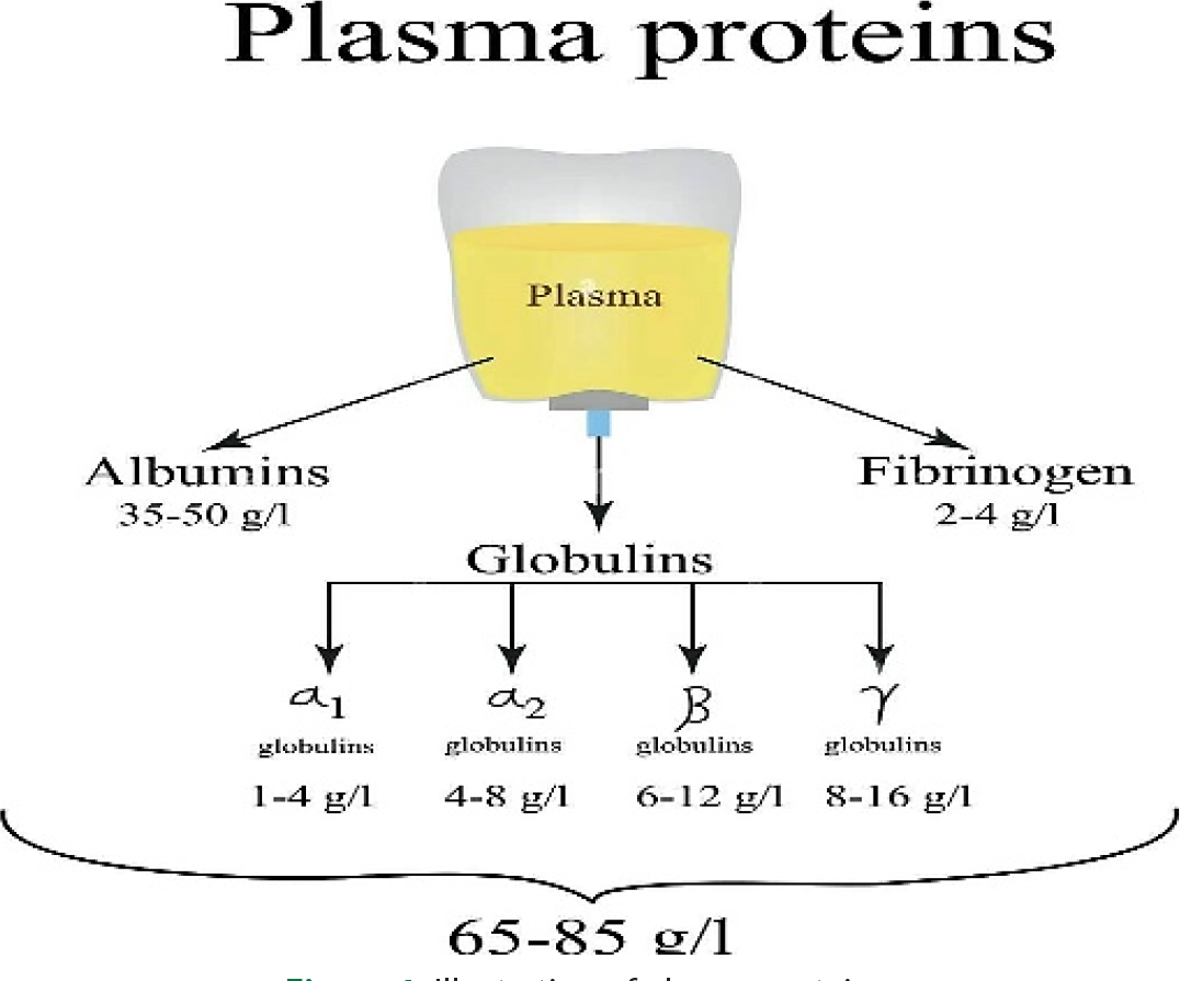
Figure 1:
Illustration of plasma proteins.
CONCLUSION
In this paper a statistical report has been presented on plasma Proteins and Clinical Significance plasma proteins play a dominant role in the pathogenesis of a variety of diseases and clinical syndromes (O’Reilly, 1980). The names of some proteins have become the names of diseases; the specific items discussed in this paper include albumin, fibrinogen, globulins, liver disease, pregnancy, infancy and old age, haemorrhagic disorders, amyloidosis, and other topics (Stewart et al., 1991). The treatment of these complex subjects is aimed at the person without specialized education Separating serum proteins by electrophoresis is a valuable diagnostic tool as well as a way to monitor clinical progress (McElnay and D’Arcy, 1983). Current research regarding blood plasma proteins is centred on performing proteomics analyses of serum/plasma in the search for biomarkers. It is observed in this study Blood proteins, also termed plasma proteins, are proteins present in blood plasma. They serve many different functions, including transport of lipids, hormones, vitamins and minerals in activity and functioning of the immune system. Other blood proteins act as enzymes, complement components, protease inhibitors or kinin precursors. Contrary to popular belief, haemoglobin is not a blood protein, as it is carried within red blood cells, rather than in the blood serum.
Acknowledgments
None.
Conflict of Interest
The author declares that there is no conflict of interest.
References
Aarons, L. J., Rowland, M.. (1981). Kinetics of drug displacement interactions. Journal of Pharmacokinetics and Biopharmaceutics, 9(2), 181-190. https://doi.org/10.1007/BF01068081
Aggeler, P. M., O’Reilly, R. A., Leong, L., Kowitz, P. E.. (1967). Potentiation of anticoagulant effect of warfarin by phenylbutazone. New England Journal of Medicine, 276(9), 496-501. https://doi.org/10.1056/NEJM196703022760904
Banfield, C., O’Reilly, R., Chan, E., Rowland, M.. (1983). Phenylbutazone-warfarin interaction in man: Further stereochemical and metabolic considerations. British Journal of Clinical Pharmacology, 16(6), 669-675. https://doi.org/10.1111/j.1365-2125.1983.tb02239.x
Bjornsson, T. D., Meffin, P. J., Swezey, S., Blaschke, T. F.. (1979a September). Clofibrate displaces warfarin from plasma proteins in man: An example of a pure displacement interaction. Journal of Pharmacology and Experimental Therapeutics, 210(3), 316-321.
Bjornsson, T. D., Meffin, P. J., Swezey, S. E., Blaschke, T. F.. (1979b September). Effects of clofibrate and warfarin alone and in combination on the disposition of vitamin K1. Journal of Pharmacology and Experimental Therapeutics, 210(3), 322-326. https://doi.org/10.1016/S0022-3565(25)31755-6
Brodie, B. B.. (1965). Displacement of one drug by another from carrier or receptor sites. Proceedings of the Royal Society of Medicine, 58(11 Part 2 Pt. 2), 946-955. https://doi.org/10.1177/003591576505811P202
Christensen, L. K., Hansen, J. M., Kristensen, M.. (1963). Sulphaphenazole-induced hypoglycaemic attacks in tolbutamide-treated diabetics. Lancet, 2(7321), 1298-1301. https://doi.org/10.1016/S0140-6736(63)90847-X
Faed, E. M.. (1981). Protein Binding of Drugs in plasma, interstitial Fluid and tissues: Effect on pharmacokinetics. European Journal of Clinical Pharmacology, 21(1), 77-81. https://doi.org/10.1007/BF00609592
Fox, S. L.. (1964). Potentiation of anticoagulants caused by pyrazole compounds. JAMA, 188, 320-321. https://doi.org/10.1001/Jama.1964.03060290124046
Gibaldi, M., Levy, G., McNamara, P. J.. (1978). Effect of plasma protein and tissue binding on the biologic half-life of drugs. Clinical Pharmacology and Therapeutics, 24(1), 1-4. https://doi.org/10.1002/cpt19782411
Griner, P. F., Raisz, L. G., Rickles, F. R., Wiesner, P. J., Odoroff, C. L.. (1971). Chloral hydrate and warfarin interaction: Clinical significance. Annals of Internal Medicine, 74(4), 540-543. https://doi.org/10.7326/0003-4819-74-4-540
Hansen, J. M., Christensen, L. K.. (1977). Drug interactions with oral sulphonylurea hypoglycaemic drugs. Drugs, 13(1), 24-34. https://doi.org/10.2165/00003495-197713010-00003
Koch-Weser, J., Sellers, E. M.. (1976). Binding of drugs to serum albumin (first of two parts). New England Journal of Medicine, 294(6), 311-316. https://doi.org/10.1056/NEJM197602052940605
Liegler, D. G., Henderson, E. S., Hahn, M. A., Oliverio, V. T.. (1969 November-December). The effect of organic acids on renal clearance of methotrexate in man. Clinical Pharmacology and Therapeutics, 10(6), 849-857. https://doi.org/10.1002/cpt1969106849
MacKichan, J. J.. (1984). Pharmacokinetic consequences of drug displacement from blood and tissue proteins. Clinical Pharmacokinetics, 9(Suppl. 1), 32-41. https://doi.org/10.2165/00003088-198400091-00005
McElnay, J. C., D’Arcy, P. F.. (1983). Protein binding displacement interactions and their clinical importance. Drugs, 25(5), 495-513. https://doi.org/10.2165/00003495-198325050-00003
O’Reilly, R. A.. (1980). Stereoselective interaction of trimethoprim-sulfamethoxazole with the separated enantiomorphs of racemic warfarin in man. New England Journal of Medicine, 302(1), 33-35. https://doi.org/10.1056/NEJM198001033020106
O’Reilly, R. A., Goulart, D. A.. (1981). Comparative interaction of sulfinpyrazone and phenylbutazone with racemic warfarin: Alteration in vivo of free fraction of plasma warfarin. Journal of Pharmacology and Experimental Therapeutics, 219(3), 691-694. https://doi.org/10.1016/S0022-3565(25)32863-6
O’Reilly, R. A., Trager, W. F., Motley, C. H., Howald, W.. (1980). Stereoselective interaction of phenylbutazone with [12C/13C]warfarin pseudoracemates in man. Journal of Clinical Investigation, 65(3), 746-753. https://doi.org/10.1172/JCI109722
Perucca, E., Hebdige, S., Frigo, G. M., Gatti, G., Lecchini, S., Crema, A.. (1980). Interaction between phenytoin and valproic acid: Plasma protein binding and metabolic effects. Clinical Pharmacology and Therapeutics, 28(6), 779-789. https://doi.org/10.1038/clpt.1980.235
Pond, S. M., Birkett, D. J., Wade, D. N.. (1977). Mechanisms of inhibition of tolbutamide metabolism: Phenylbutazone, oxyphenbutazone, sulfaphenazole. Clinical Pharmacology and Therapeutics, 22(5 Pt. 1), 573-579. https://doi.org/10.1002/cpt1977225part1573
Sellers, E. M.. (1979). Plasma protein displacement interactions are rarely of clinical significance. Pharmacology, 18(5), 225-227. https://doi.org/10.1159/000137256
Sellers, E. M., Koch-Weser, J.. (1970). Potentiation of warfarin-induced hypoprothrombinemia by chloral hydrate. New England Journal of Medicine, 283(16), 827-831. https://doi.org/10.1056/NEJM197010152831601
Sellers, E. M., Koch-Weser, J.. (1971). Kinetics and clinical importance of displacement of warfarin from albumin by acidic drugs. Annals of the New York Academy of Sciences, 179, 213-225. https://doi.org/10.1111/j.1749-6632.1971.tb46901.x
Stewart, C. F., Fleming, R. A., Germain, B. F., Seleznick, M. J., Evans, W. E.. (1991). Aspirin alters methotrexate disposition in rheumatoid arthritis patients. Arthritis and Rheumatism, 34(12), 1514-1520. https://doi.org/10.1002/art.1780341207
Tillement, J. P., Zini, R., Mattei, C., Singlas, E.. (1973). Effect of phenylbutazone on the binding of vitamin K antagonists to albumin. European Journal of Clinical Pharmacology, 6(1), 15-18. https://doi.org/10.1007/BF00561795
Toon, S., Low, L. K., Gibaldi, M., Trager, W. F., O’Reilly, R. A., Motley, C. H., Goulart, D. A.. (1986). The warfarin-sulfinpyrazone interaction: Stereochemical considerations. Clinical Pharmacology and Therapeutics, 39(1), 15-24. https://doi.org/10.1038/clpt.1986.3
Wilkinson, G. R., Shand, D. G.. (1975). Commentary: A physiological approach to hepatic drug clearance. Clinical Pharmacology and Therapeutics, 18(4), 377-390. https://doi.org/10.1002/cpt1975184377