


Prostate adenocarcinoma is a common malignancy in elderly males and may present with lower urinary tract symptoms, particularly when advanced or metastatic. We report a 65-year-old male presenting with progressive urinary complaints, chronic low back pain, and generalized weakness. Laboratory evaluation revealed markedly elevated serum prostate-specific antigen levels. Transrectal ultrasound-guided biopsy confirmed prostatic adenocarcinoma with a Gleason score of 4 + 3. Advanced imaging demonstrated metastatic involvement of pelvic lymph nodes and multiple skeletal sites. Prostate-specific membrane antigen positron emission tomography/computed tomography enabled comprehensive assessment of tumor extent and confirmed disseminated disease. The patient was managed initially with symptomatic therapy and androgen deprivation therapy, followed by bilateral surgical castration after multidisciplinary evaluation. This case highlights the diagnostic challenges associated with advanced prostate carcinoma and emphasizes the clinical value of molecular imaging for accurate staging, prognostication, and therapeutic decision-making. Early recognition and individualized management remain essential to optimize outcomes in metastatic prostate cancer.
Abstract
Share on Mobile
Scan to access on mobile
Keywords
Bone Metastases
Lower Urinary Tract Symptoms
Metastatic Prostate Cancer
Prostate Adenocarcinoma
Prostate-Specific Membrane Antigen Imaging
INTRODUCTION
Prostate adenocarcinoma is one of the most frequently diagnosed malignancies among elderly males and often presents with Lower Urinary Tract Symptoms (LUTS). In advanced stages, the disease may metastasize to distant organs, particularly bone and lymph nodes, increasing morbidity and complicating diagnosis. Chronic inflammation has been implicated in prostate carcinogenesis through cytokine-mediated alterations in the tumor microenvironment that promote cellular proliferation and metastatic progression (Stark et al., 2015).
Management strategies depend on disease stage and may include androgen deprivation therapy, chemotherapy, radiation therapy, or targeted treatments. Prognosis is influenced by factors such as age, comorbid conditions, tumor grade, and metastatic burden (Leslie et al., 2023). Early detection is critical for improving outcomes, and screening tools such as digital rectal examination and prostate-specific antigen testing remain widely used in clinical practice (Drudge-Coates et al., 2018).
The etiology of prostate cancer is multifactorial, involving nonmodifiable determinants such as age, ethnicity, and genetic susceptibility, along with modifiable influences including chronic inflammation, infections, environmental exposures, and lifestyle factors (Abidi et al., 2018; Berenguer et al., 2023; Ng, 2021). Because clinical behavior varies widely, accurate staging is essential for appropriate therapeutic planning.
This report presents a case of advanced metastatic prostate adenocarcinoma in an elderly male with persistent urinary symptoms, highlighting diagnostic challenges and emphasizing the role of prostate-specific membrane antigen positron emission tomography/computed tomography in comprehensive disease staging.
CASE PRESENTATION
A 65-year-old male with no known comorbidities and no family history of malignancy presented to the Department of Nephrology at Shree Gajanan Maharaj Hospital with progressive LUTS, including increased urinary frequency, weak urinary stream, straining during micturition, and post-void dribbling for approximately one year. He also reported a burning sensation during defecation for two months, chronic low back pain for one year, and intermittent diffuse pain over the bilateral posterolateral chest wall for five months.
On general examination, the patient was clinically stable with a Karnofsky Performance Status score of 80. No peripheral lymphadenopathy or abdominal abnormalities were detected. Digital rectal examination revealed a smooth, enlarged prostate with a palpable median lobe without rectal wall fixation or mucosal irregularity.
Laboratory evaluation demonstrated a markedly elevated serum prostate-specific antigen level of 396 ng/mL (reference range <4 ng/mL). Transabdominal ultrasonography showed an enlarged prostate measuring 4.1 × 4.2 × 4.5 cm, associated with urinary bladder wall thickening and minimal post-void residual urine volume of 15 mL. Transrectal ultrasound-guided prostate biopsy confirmed prostatic adenocarcinoma with a Gleason score of 4 + 3 (Grade Group 3).
For staging, whole-body positron emission tomography/computed tomography using fluorodeoxyglucose revealed multiple sclerotic skeletal lesions that were non-avid, along with mildly enlarged perirectal and presacral lymph nodes demonstrating low-grade metabolic activity, suggestive of metastatic disease.
Subsequent gallium-68 prostate-specific membrane antigen positron emission tomography/computed tomography demonstrated intense tracer uptake throughout the prostate gland (maximum standardized uptake value 32.61), with extension toward the bladder base and seminal vesicles and loss of the intervening fat plane with the anterior rectal wall (Figure 1). Multiple prostate-specific membrane antigen-avid lymph nodes were detected in the pelvic and retroperitoneal regions, along with widespread skeletal metastases involving the skull, sternum, clavicle, vertebral column, pelvis, and ribs (Figure 2). Corresponding computed tomography images demonstrated sclerotic lesions in the thoracic spine consistent with metastatic involvement (Figure 3).
The patient was initially treated symptomatically with oral tamsulosin 0.4 mg once daily and dutasteride 0.5 mg once daily. Given the extensive metastatic burden, androgen deprivation therapy was initiated with oral bicalutamide 50 mg three times daily along with calcium supplementation, as per institutional clinical protocol. After multidisciplinary discussion regarding therapeutic options, the patient elected to undergo bilateral orchiectomy as definitive hormonal therapy.
The diagnosis of metastatic prostate adenocarcinoma with nodal and skeletal involvement was established based on histopathology and advanced molecular imaging. The patient remains under regular follow-up with serial prostate-specific antigen monitoring and clinical evaluation to assess treatment response and detect disease progression.
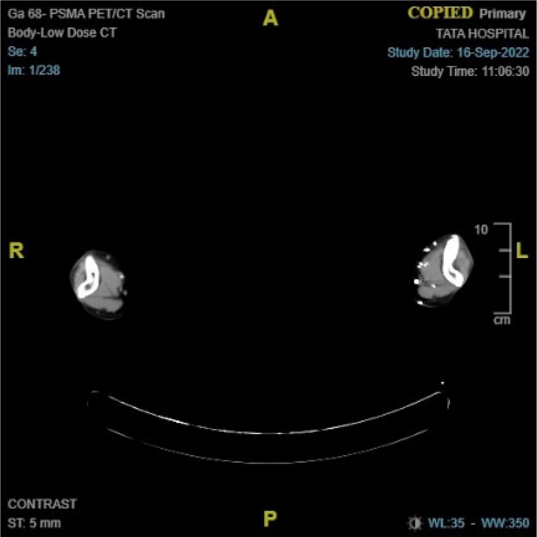
Figure 1:
Axial computed tomography image showing prostatic enlargement with loss of the fat plane between the prostate and anterior rectal wall.
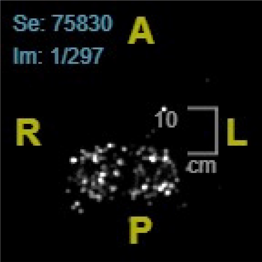
Figure 2:
Whole-body gallium-68 prostate-specific membrane antigen positron emission tomography maximum intensity projection image demonstrating intense tracer uptake in the prostate with widespread skeletal metastases.
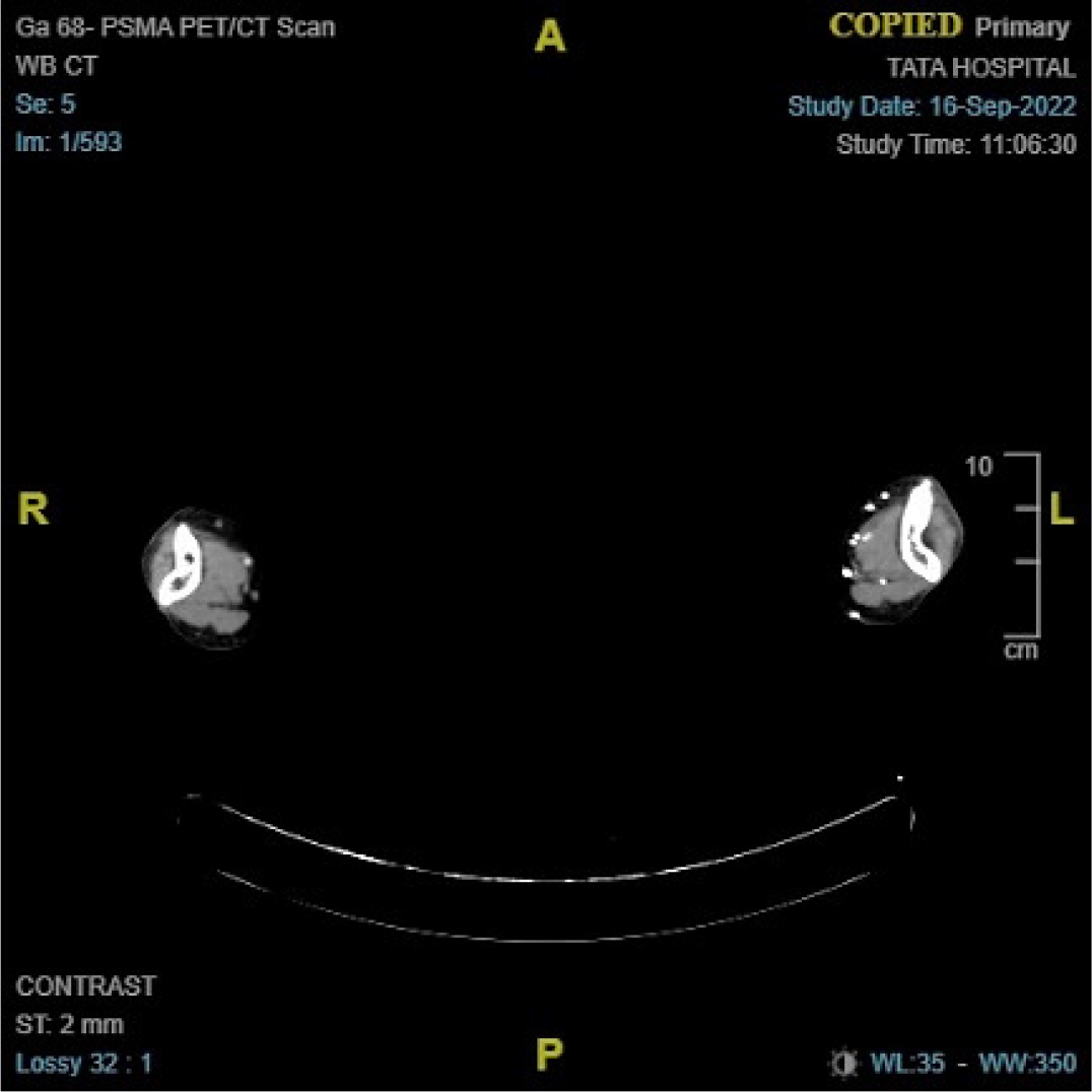
Figure 3:
Computed tomography image showing sclerotic thoracic vertebral lesions consistent with metastatic involvement.
DISCUSSION
This case highlights the diagnostic challenges associated with metastatic prostate adenocarcinoma in elderly males presenting with persistent LUTS. Such symptoms are frequently attributed to benign prostatic hyperplasia, which may delay suspicion of malignancy, particularly when systemic warning signs are absent. The markedly elevated prostate-specific antigen level in this patient prompted further investigation, underscoring the importance of biochemical screening in directing diagnostic evaluation (Descotes, 2019; Drudge-Coates et al., 2018; Ng, 2021).
Advanced molecular imaging played a decisive role in accurate staging. Although initial positron emission tomography/computed tomography demonstrated skeletal abnormalities suggestive of metastatic disease, gallium-68 prostate-specific membrane antigen positron emission tomography/computed tomography provided superior lesion characterization and a more comprehensive assessment of nodal and skeletal involvement. Prostate-specific membrane antigen-based imaging has demonstrated higher sensitivity and specificity than conventional imaging modalities, particularly for detecting metastatic lesions at low tumor burden, thereby improving staging accuracy and influencing therapeutic planning (Crowley et al., 2021; Nakazawa et al., 2017; Yu et al., 2023). Accurate staging through such imaging directly influences therapeutic decision-making and prognostication (Descotes, 2019).
Androgen deprivation therapy remains the cornerstone of management for metastatic prostate cancer. In the present case, the patient elected bilateral surgical orchiectomy after detailed counseling regarding available therapeutic options. Surgical castration provides rapid and sustained testosterone suppression, eliminates adherence concerns, and may represent a cost-effective strategy in appropriately selected individuals. Selection of an androgen deprivation modality should consider disease burden, patient preference, expected compliance, and healthcare resource availability (Crowley et al., 2021; Nakazawa et al., 2017; Yu et al., 2023).
From a pharmacy practice perspective, this case emphasizes the integral role of pharmacists in multidisciplinary oncology care. Pharmacists contribute to therapeutic optimization through medication counseling, monitoring for adverse drug reactions, evaluating drug-drug interactions, ensuring adherence to hormonal therapy, and educating patients regarding long-term treatment implications. Such collaborative involvement is essential for improving treatment outcomes and quality of life in patients with advanced prostate cancer (Rawla, 2019).
This report is limited by its single-patient design and lack of long-term follow-up. Nevertheless, carefully documented case reports provide clinically relevant insights, particularly regarding real-world diagnostic decision-making and individualized treatment strategies. Continued reporting of similar cases may support refinement of diagnostic algorithms and management approaches for metastatic prostate adenocarcinoma (Gandaglia et al., 2021; Porcacchia et al., 2022; Rawla, 2019).
CONCLUSION
This case emphasizes the importance of comprehensive diagnostic evaluation in elderly males presenting with persistent LUTS, as these manifestations may represent early indicators of advanced prostate adenocarcinoma. Prompt consideration of malignancy, supported by appropriate biochemical testing and targeted imaging, is essential to minimize diagnostic delay and facilitate timely management.
The use of prostate-specific membrane antigen positron emission tomography/computed tomography enabled accurate staging and directly informed therapeutic decision-making in this patient. Management of metastatic prostate cancer requires an individualized, patient-centered approach integrating disease burden, patient preferences, and multidisciplinary expertise. Documentation of clinically significant cases such as this contributes to improved diagnostic awareness and supports refinement of management strategies for advanced prostate cancer.
Acknowledgments
The authors thank the patient for consenting to the publication of the clinical details of this case.
Conflict of Interest
The authors declare that there is no conflict of interest.
Ethics Statement
The authors certify that they have obtained all appropriate patient consent.
References
Abidi, S. H., Bilwani, F., Ghias, K., Abbas, F.. (2018). Viral etiology of prostate cancer: Genetic alterations and immune response. International Journal of Surgery,, 52, 136-140. https://doi.org/10.1016/j.ijsu.2018.02.050
Berenguer, C. V., Pereira, F., Câmara, J. S., Pereira, J. A. M.. (2023). Underlying features of prostate cancer: Statistics, risk factors, and emerging methods for its diagnosis. Current Oncology,, 30(2), 2300-2321. https://doi.org/10.3390/curroncol30020178
Crowley, F., Sterpi, M., Buckley, C., Margetich, L., Handa, S., Dovey, Z.. (2021). A review of the pathophysiological mechanisms underlying castration-resistant prostate cancer. Research and Reports in Urology,, 13, 457-472. https://doi.org/10.2147/RRU.S264722
Descotes, J. L.. (2019). Diagnosis of prostate cancer. Asian Journal of Urology,, 6(2), 129-136. https://doi.org/10.1016/j.ajur.2018.11.007
Drudge-Coates, L., Oh, W. K., Tombal, B.. (2018). Recognizing symptom burden in advanced prostate cancer: A global patient and caregiver survey. Clinical Genitourinary Cancer,, 16(2), e411-e419. https://doi.org/10.1016/j.clgc.2017.09.015
Gandaglia, G., Leni, R., Bray, F.. (2021). Epidemiology and prevention of prostate cancer. European Urology Oncology,, 4(6), 877-892. https://doi.org/10.1016/j.euo.2021.09.006
Leslie, S. W., Soon-Sutton, T. L., Ria, R. I., Sajjad, H., Skelton, W. P.. (2023). In StatPearls.
Nakazawa, M., Paller, C., Kyprianou, N.. (2017). Mechanisms of therapeutic resistance in prostate cancer. Current Oncology Reports,, 19(2), 13. https://doi.org/10.1007/s11912-017-0568-7
Ng, K. L.. (2021). In Prostate cancer, 17-27).
Porcacchia, A. S., Pires, G. N., Ortiz, V., Andersen, M. L., Tufik, S.. (2022). Prostate cancer mortality and costs of prostate surgical procedures in the Brazilian public health system. International Brazilian Journal of Urology,, 48(3), 583-590. https://doi.org/10.1590/S1677-5538.IBJU.2021.0781
Rawla, P.. (2019). Epidemiology of prostate cancer. World Journal of Oncology,, 10(2), 63-89. https://doi.org/10.14740/wjon1191
Stark, T., Livas, L., Kyprianou, N.. (2015). Inflammation in prostate cancer progression and therapeutic targeting. Translational Andrology and Urology,, 4(4), 455-463. https://doi.org/10.3978/j.issn.2223-4683.2015.04.12
Yu, E. M., Hwang, M. W., Aragon-Ching, J. B.. (2023). Mechanistic insights on localized to metastatic prostate cancer transition and therapeutic opportunities. Research and Reports in Urology,, 15, 519-529. https://doi.org/10.2147/RRU.S386517